What is hydrocephalus?
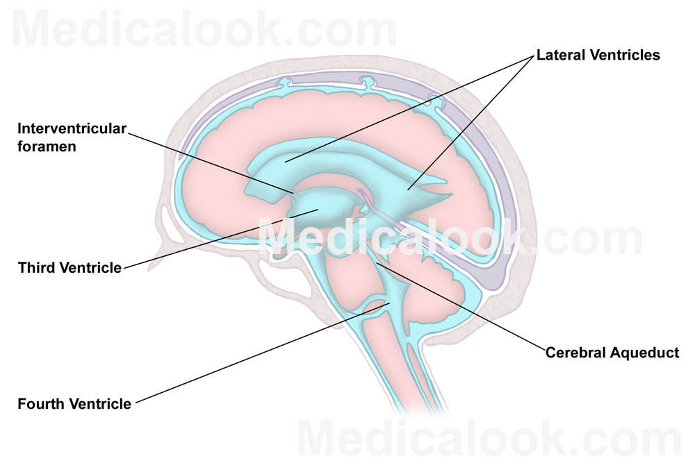
Hydrocephalus is a disorder of the brain that results in a build up of fluid (cerebrospinal fluid or CSF) that eventually damages the brain. In young children, the head enlarges as the fluid chambers of the brain, know as ventricles , become larger.
 This damages the brain and impairs the child’s development. Babies feed poorly, are irritable, vomit and fail to develop basic motor or cognitive skills. In older children and adults the enlarging ventricles cause pressure in the brain. This results in headaches, vomiting, crossed eyes, poor balance, and eventually lethargy and death.
What causes hydrocephalus?
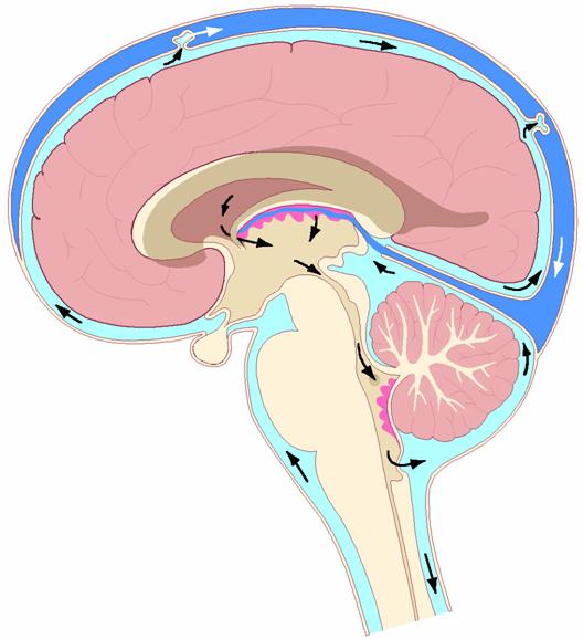
Hydrocephalus is caused by an imbalance between the rate of CSF production and its absorption. The brain makes CSF at a relatively constant rate, mostly within the ventricles. The fluid flows through the ventricles of the brain, exits the base of brain and is then reabsorbed back into the blood stream. The build up of CSF in hydrocephalus is nearly always caused by a blockage to the flow of CSF within the brain or an impairment of the CSF absorption pathways outside the brain. Some of the blockages are structural; such as a scarring in the narrow fluid channel between the 3rd and 4th ventricle (know as aqueduct stenosis), a tumor or a developmental abnormality.

The arrows show the direction of CSF from where it formed, in the ventricles, to where it is absorbed outside the brain. (From:http://www.control.tfe.umu.se/Ian/CSF/)
Infections of the brain, particularly when they occur early in life, can cause loss of the brain’s ability to absorb CSF. This can be the result of bacterial, viral or parasitic infections, which are a significant problem in developing countries. These infections are frequently contracted around the time of birth, and although uncommon in the developed world, cause at least half of the hydrocephalus cases treated by our team in Haiti.
How is hydrocephalus diagnosed?
In infants and children under the age of two years the gradual build up of fluid results in excessive head growth. The head appears much larger then would be normal for the child’s age when compared to normal children. In these children the diagnosis can be made easily by measuring the head circumference and plotting the size against the age on a growth curve . These children will have a large soft spot (fontanel), which is usually tense. The scalp veins may appear more pronounced and the eyes may appear to be ‘setting’ on the lower lid. An ultrasound can be used to see the size of the fluid spaces through the open fontanel.
In older children and adults, or if a better view of the structures of the brain is required, a CT scan or MRI scan is needed. CT scans use x-rays and a computer to reconstruct images of the brain in ‘slices’. MRI scans by contrast create images of the brain based on the magnetic properties of water molecules and do not require x-rays. CT scanners are uncommon in the developing world because of the cost and complex technology required. MRI machines, which are significantly more complicated and expensive then CT scanners, are virtually unavailable. We have access to both ultrasound and CT imaging at the hospital for all of our children because of the generous donation of equipment to our program and the HBMPM.
How is hydrocephalus treated?
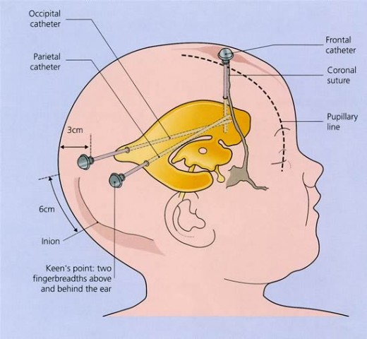
Reestablishing the balance between CSF production and absorption is the best way to treat hydrocephalus. Practically, this usually involves an operation to allow CSF to escape the brain where the fluid can be reabsorbed or by placing a tube to divert to fluid away from the brain.
These diversion tubes, or shunts, are prone to blockage, or can become infected. Children with shunts frequently require additional operations for replacement, removal or repair of their shunt because of malfunction or infection. In spite of these drawbacks, shunts are sometimes the only option to treat a child’s hydrocephalus.
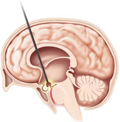
The preferred approach to treat hydrocephalus avoids leaving a shunt tube in the child. This is accomplished by making a small opening in the base of the 3rd ventricle with the aide of a camera (called an endoscopic third ventriculostomy or ETV).
When anatomically feasible, this operation allows CSF to exit the brain and be reabsorbed through the normal CSF path. Sometimes this is not possible because of scarring, either inside the ventricles or outside the brain in the CSF spaces. To improve the success of an ETV, CSF production can be reduced at it’s source, by cauterizing the choroid plexus.
-Dr. John Ragheb MD


